Home

Flamme Knospe Prägnant protein mikroskop Süd Klappe heimlich

Supermicroscope for Protein Crystals - Universität Hamburg

Imaging proteins at the single-molecule level | PNAS

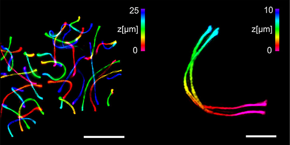
Innenrücktitelbild: Particle‐by‐Particle In Situ Characterization of the  Protein Corona via Real‐Time 3D Single‐Particle‐Tracking Spectroscopy  (Angew. Chem. 41/2021) - Tan - 2021 - Angewandte Chemie - Wiley Online  Library
Innenrücktitelbild: Particle‐by‐Particle In Situ Characterization of the Protein Corona via Real‐Time 3D Single‐Particle‐Tracking Spectroscopy (Angew. Chem. 41/2021) - Tan - 2021 - Angewandte Chemie - Wiley Online Library

Microscopy seminars | Cells in Motion

Electron micrographs of the spike protein of Sars-CoV-2 - YouTube

Cryo Electron Microscope | Krios G4 Cryo-TEM | Thermo Fisher Scientific - US

Light microscopy provides a deep look into protein structure ›  Friedrich-Alexander-Universität Erlangen-Nürnberg
Light microscopy provides a deep look into protein structure › Friedrich-Alexander-Universität Erlangen-Nürnberg

Motor proteins caught “swinging on monkey bars” - YouTube

Inhibition of Bax Channel-Forming Activity by Bcl-2

Unter dem Mikroskop - tierwelt.ch | TierWelt

Verirrte Proteine – KlarText

Inside or outside? A new collection of Gateway vectors allowing plant  protein subcellular localization or over-expression - ScienceDirect
Inside or outside? A new collection of Gateway vectors allowing plant protein subcellular localization or over-expression - ScienceDirect

Researchers Map Structure of Coronavirus "Spike" Protein - Scientific  American
Researchers Map Structure of Coronavirus "Spike" Protein - Scientific American

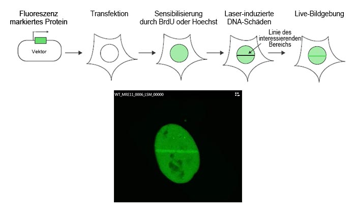
Visualisierung von DNA-Reparatur-Proteinen mit dem konfokalen Mikroskop  FV3000 | Olympus LS
Visualisierung von DNA-Reparatur-Proteinen mit dem konfokalen Mikroskop FV3000 | Olympus LS

Imaging proteins at the single-molecule level | PNAS

Top-Methode: Proteine atomgenau abbilden - science.ORF.at

Under the Electron Microscope – A 3-D Image of an Individual Protein -  Berkeley Lab
Under the Electron Microscope – A 3-D Image of an Individual Protein - Berkeley Lab

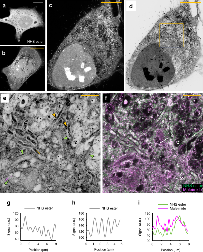
Light microscopy of proteins in their ultrastructural context | Nature  Communications
Light microscopy of proteins in their ultrastructural context | Nature Communications

High-end Microscopy Refined - Chair of Biotechnology

Der Computer als Mikroskop - Newsportal - Ruhr-Universität Bochum

New Light Microscope Can View Protein Arrangement in Cell Structures |  National Institutes of Health (NIH)
New Light Microscope Can View Protein Arrangement in Cell Structures | National Institutes of Health (NIH)
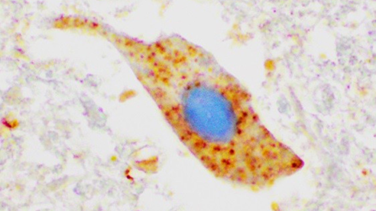
ALS disease mechanism discovered

Texas A&M researchers discover COVID-19 mechanism that helps it escape  immune system
Texas A&M researchers discover COVID-19 mechanism that helps it escape immune system

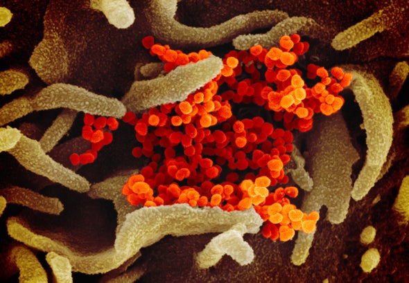
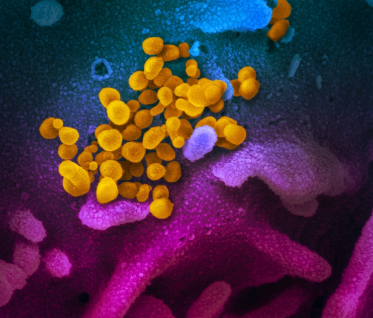
New Images of Novel Coronavirus SARS-CoV-2 Now Available | NIH: National  Institute of Allergy and Infectious Diseases
New Images of Novel Coronavirus SARS-CoV-2 Now Available | NIH: National Institute of Allergy and Infectious Diseases

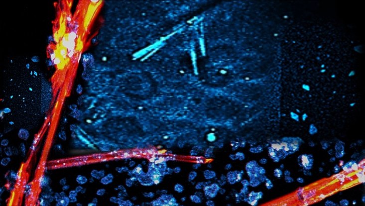
An Introduction to the Light Microscope, Light Microscopy Techniques and  Applications | Technology Networks
An Introduction to the Light Microscope, Light Microscopy Techniques and Applications | Technology Networks

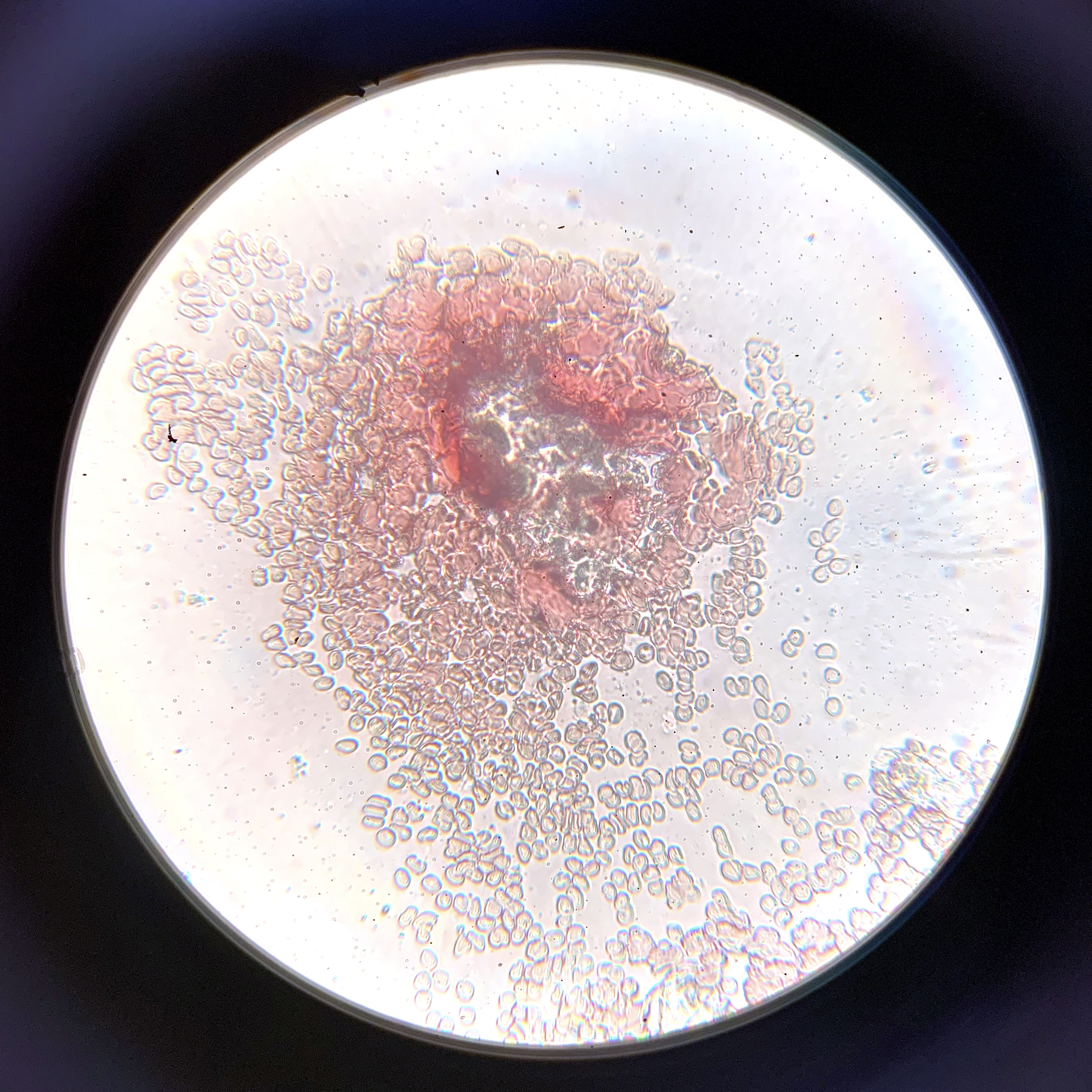
Under the Microscope: Blood | Office for Science and Society - McGill  University
Under the Microscope: Blood | Office for Science and Society - McGill University